Prof. Dr. Christian Stigloher
Diving into nervous systems with electron microscopy tomography
Institute: Biocenter, Imaging Core Facility
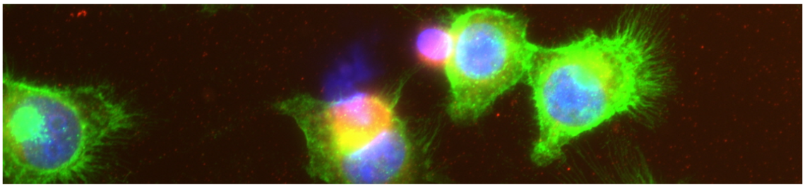
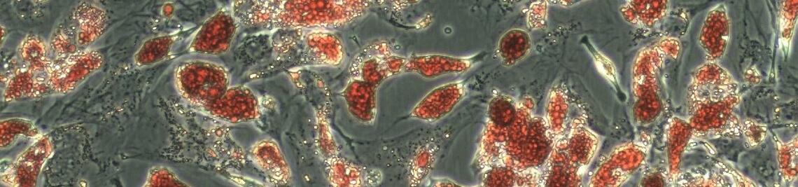
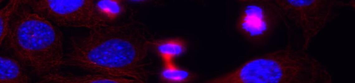
Abstract: We apply electron tomography as high 3D resolution imaging technology to study synaptic architecture and use a combination of two models where most appropriate: The C. elegans nervous system for efficient candidate identification and manipulation and the nervous system of the zebrafish larva as vertebrate model to allow a view on evolutionary conservation. Furthermore, we use correlated light and electron microscopy (CLEM) approach combining the advantages of both techniques.
Chair: Karen Auweiler