Visualizing the Synaptic and Cellular Ultrastructure in Neurons Differentiated from Human Induced Neural Stem Cells—An Optimized Protocol
02.03.2020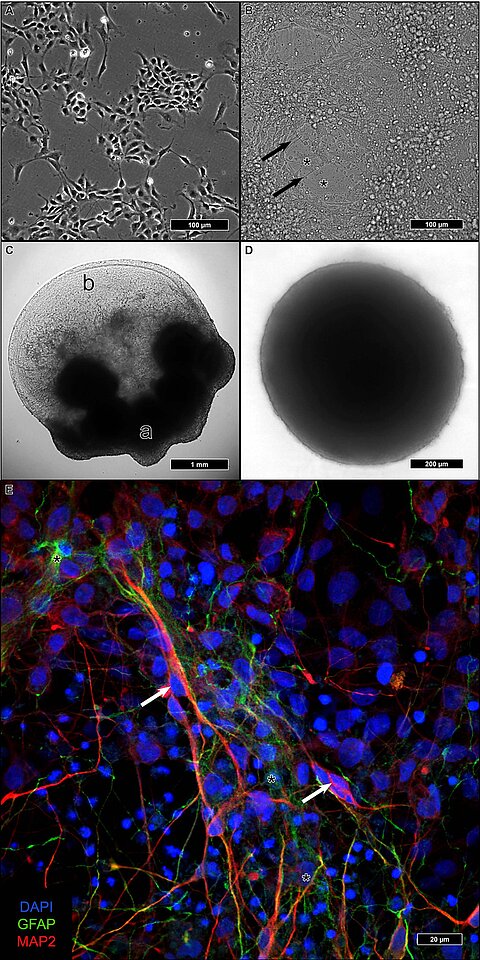
Visualizing the Synaptic and Cellular Ultrastructure in Neurons Di erentiated from Human Induced Neural Stem Cells—An Optimized Protocol
Philipp Capetian, Lorenz Müller, Jens Volkmann, Manfred Heckmann, Süleyman Ergün and NicoleWagner
Int. J. Mol. Sci. 2020, 21(5), 1708
The size of the synaptic subcomponents falls below the limits of visible light microscopy. Despite new developments in advanced microscopy techniques, the resolution of transmission electron microscopy (TEM) remains unsurpassed. The requirements of tissue preservation are very high, and human post mortem material often does not offer adequate quality. However, new reprogramming techniques that generate human neurons in vitro provide samples that can easily fulfill these requirements. The objective of this study was to identify the culture technique with the best ultrastructural preservation in combination with the best embedding and contrasting technique for visualizing neuronal elements. Two induced neural stem cell lines derived from healthy control subjects underwent differentiation either adherent on glass coverslips, embedded in a droplet of highly concentrated Matrigel, or as a compact neurosphere. Afterward, they were fixed using a combination of glutaraldehyde (GA) and paraformaldehyde (PFA) followed by three approaches (standard stain, Ruthenium red stain, high contrast en-bloc stain) using different combinations of membrane enhancing and contrasting steps before ultrathin sectioning and imaging by TEM. The compact free-floating neurospheres exhibited the best ultrastructural preservation. High-contrast en-bloc stain offered particularly sharp staining of membrane structures and the highest quality visualization of neuronal structures. In conclusion, compact neurospheres growing under free-floating conditions in combination with a high contrast en-bloc staining protocol, offer the optimal preservation and contrast with a particular focus on visualizing membrane structures as required for analyzing synaptic structures.