Using Expansion Microscopy to Visualize and Characterize the Morphology of Mitochondrial Cristae
15.07.2020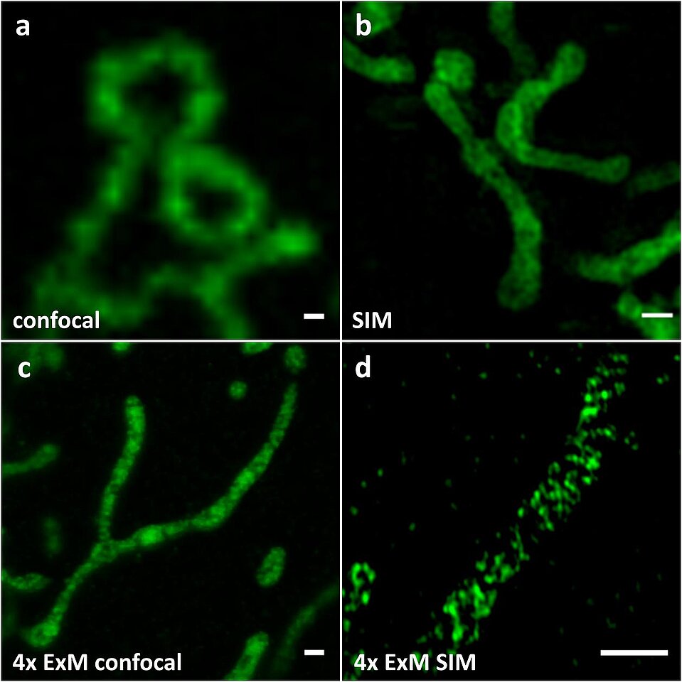
Using Expansion Microscopy to Visualize and Characterize the Morphology of Mitochondrial Cristae
Tobias C Kunz, Ralph Götz, Shiqiang Gao, Markus Sauer, Vera Kozjak-Pavlovic
Front. Cell Dev. Biol., 8:617
Mitochondria are double membrane bound organelles indispensable for biological processes such as apoptosis, cell signaling, and the production of many important metabolites, which includes ATP that is generated during the process known as oxidative phosphorylation (OXPHOS). The inner membrane contains folds called cristae, which increase the membrane surface and thus the amount of membrane-bound proteins necessary for the OXPHOS. These folds have been of great interest not only because of their importance for energy conversion, but also because changes in morphology have been linked to a broad range of diseases from cancer, diabetes, neurodegenerative diseases, to aging and infection. With a distance between opposing cristae membranes often below 100 nm, conventional fluorescence imaging cannot provide a resolution sufficient for resolving these structures. For this reason, various highly specialized super-resolution methods including dSTORM, PALM, STED, and SIM have been applied for cristae visualization. Expansion Microscopy (ExM) offers the possibility to perform super-resolution microscopy on conventional confocal microscopes by embedding the sample into a swellable hydrogel that is isotropically expanded by a factor of 4-4.5, improving the resolution to 60-70 nm on conventional confocal microscopes, which can be further increased to ∼ 30 nm laterally using SIM. Here, we demonstrate that the expression of the mitochondrial creatine kinase MtCK linked to marker protein GFP (MtCK-GFP), which localizes to the space between the outer and the inner mitochondrial membrane, can be used as a cristae marker. Applying ExM on mitochondria labeled with this construct enables visualization of morphological changes of cristae and localization studies of mitochondrial proteins relative to cristae without the need for specialized setups. For the first time we present the combination of specific mitochondrial intermembrane space labeling and ExM as a tool for studying internal structure of mitochondria.