Targeted volumetric single-molecule localization microscopy of defined presynaptic structures in brain sections
25.03.2021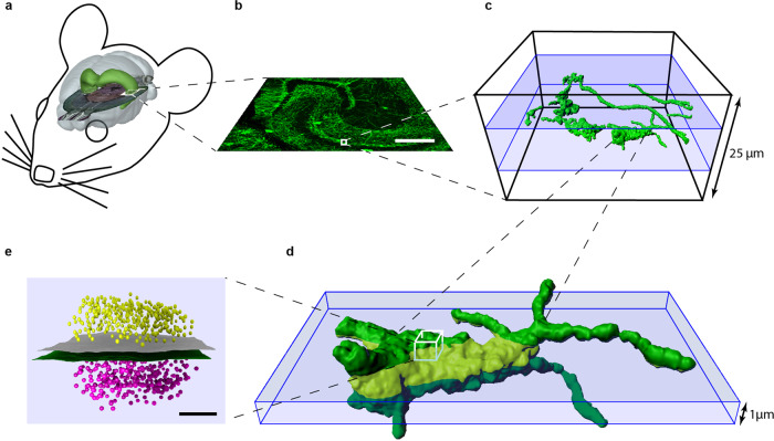
Targeted volumetric single-molecule localization microscopy of defined presynaptic structures in brain sections
Martin Pauli, Mila M. Paul, Sven Proppert, Achmed Mrestani, Marzieh Sharifi, Felix Repp, Lydia Kürzinger, Philip Kollmannsberger, Markus Sauer, Manfred Heckmann, Anna-Leena Sirén
Commun. Biol. , 4:407
Revealing the molecular organization of anatomically precisely defined brain regions is necessary for refined understanding of synaptic plasticity. Although three-dimensional (3D) single-molecule localization microscopy can provide the required resolution, imaging more than a few micrometers deep into tissue remains challenging. To quantify presynaptic active zones (AZ) of entire, large, conditional detonator hippocampal mossy fiber (MF) boutons with diameters as large as 10 μm, we developed a method for targeted volumetric direct stochastic optical reconstruction microscopy (dSTORM). An optimized protocol for fast repeated axial scanning and efficient sequential labeling of the AZ scaffold Bassoon and membrane bound GFP with Alexa Fluor 647 enabled 3D-dSTORM imaging of 25 μm thick mouse brain sections and assignment of AZs to specific neuronal substructures. Quantitative data analysis revealed large differences in Bassoon cluster size and density for distinct hip- pocampal regions with largest clusters in MF boutons.