Single-Molecule Localization Microscopy of Presynaptic Active Zones in Drosophila melanogaster after Rapid Cryofixation
21.01.2023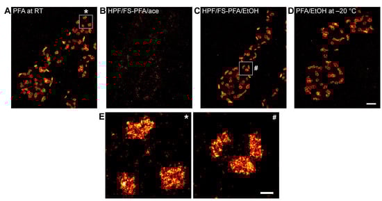
Single-Molecule Localization Microscopy of Presynaptic Active Zones in Drosophila melanogaster after Rapid Cryofixation
Achmed Mrestani, Katharina Lichter, Anna-Leena Sirén, Manfred Heckmann, Mila M. Paul and Martin Pauli
Int. J. Mol. Sci. 2023, 24(3), 2128
Single-molecule localization microscopy (SMLM) greatly advances structural studies of diverse biological tissues. For example, presynaptic active zone (AZ) nanotopology is resolved in increasing detail. Immunofluorescence imaging of AZ proteins usually relies on epitope preservation using aldehyde-based immunocompetent fixation. Cryofixation techniques, such as high-pressure freezing (HPF) and freeze substitution (FS), are widely used for ultrastructural studies of presynaptic architecture in electron microscopy (EM). HPF/FS demonstrated nearer-to-native preservation of AZ ultrastructure, e.g., by facilitating single filamentous structures. Here, we present a protocol combin- ing the advantages of HPF/FS and direct stochastic optical reconstruction microscopy (dSTORM) to quantify nanotopology of the AZ scaffold protein Bruchpilot (Brp) at neuromuscular junctions (NMJs) of Drosophila melanogaster. Using this standardized model, we tested for preservation of Brp clusters in different FS protocols compared to classical aldehyde fixation. In HPF/FS samples, presynaptic boutons were structurally well preserved with ~22% smaller Brp clusters that allowed quantifica- tion of subcluster topology. In summary, we established a standardized near-to-native preparation and immunohistochemistry protocol for SMLM analyses of AZ protein clusters in a defined model synapse. Our protocol could be adapted to study protein arrangements at single-molecule resolution in other intact tissue preparations.