Advancing Array Tomography to Study the Fine Ultrastructure of Identified Neurons in Zebrafish (Danio rerio)
26.08.2020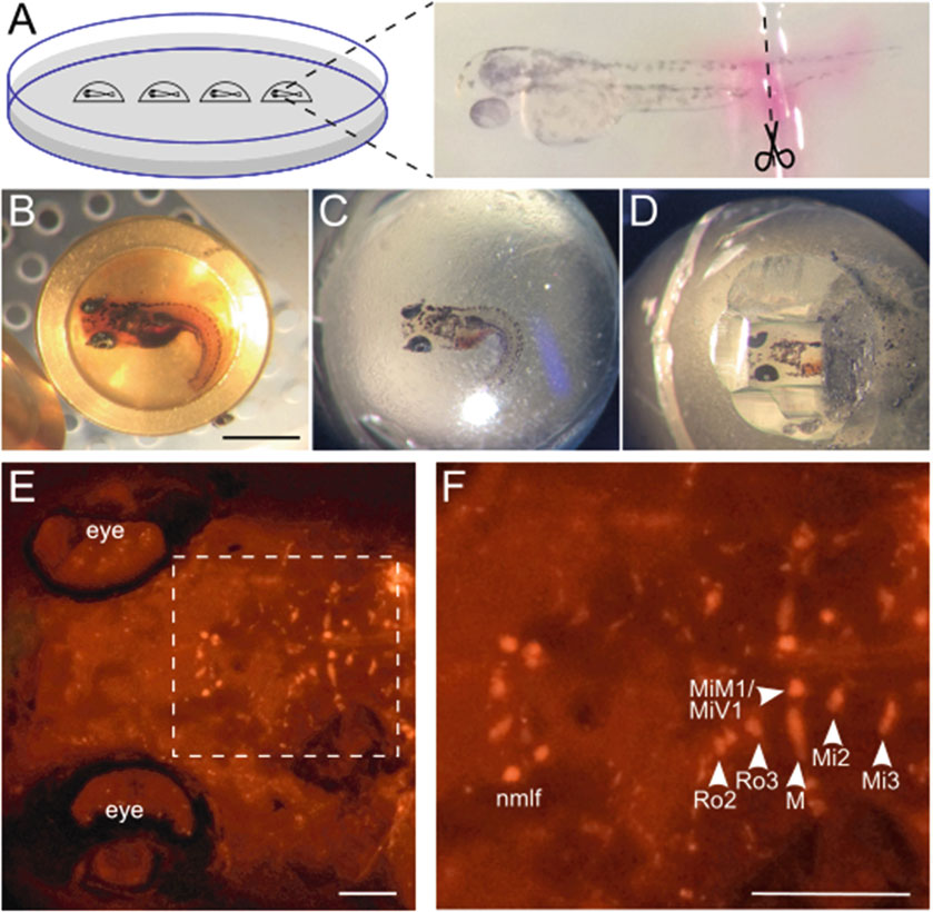
Advancing Array Tomography to Study the Fine Ultrastructure of Identified Neurons in Zebrafish (Danio rerio)
Marlene Strobel, Frederik Helmprobst, Martin Pauli, Manfred Heckmann, Christina Lillesaar, and Christian Stigloher
Springer, Volume Microscopy. Neuromethods, vol 155: 59-78
Array tomography (AT) provides a versatile workflow for correlated light and electron microscopy (CLEM). In short, biological tissues are embedded in EM-resins for immunolabeling, cut in ultrathin section arrays, which are mounted on glass slides, labeled and imaged for immunofluorescence at the light microscope and then prepared for scanning electron microscopy (SEM) imaging. Light- and electron micrographs obtained from the identical regions of interest of the same sections are then correlated to an aligned composite image series.We adapted this protocol to identify and image theMauthner neuron of the developing zebrafish embryo. The Mauthner neuron is an identifiable neuron, which can be easily labeled by retrograde tracing with for example rhodamine dextran. We take advantage of the fact that the fluorescence of rhodamine is retained after embedding in the LR White resin. Furthermore, we expanded the workflow to reach a near-to-native ultrastructural preservation and good antigenicity of the nervous tissue, by applying high pressure freezing and freeze substitution.Moreover, we add structured illumination microcopy (SIM) as imaging modality to allow tracing of fine neuronal projections and increase correlation accuracy.